 Send a Whatsapp Message
 Free shipping on orders above £49 |
Auscultation is a clinical skill worth learning. The ability to accurately identify various heart, lung, and bowel sounds can help you quickly and efficiently assess patients and monitor their condition. The following conditions and equipment will help you perform a proper auscultation:
-
- A quiet, well-lit, warm room.
- An appropriately disrobed patient. This allows you to place the stethoscope directly on the chest or back and will eliminate distortions and frictional noise from clothing.
- The ability to examine the patient supine, sitting, and in left lateral recumbent positions. You may hear different sounds, especially abnormal ones, in different positions.
- A stethoscope with both a bell and diaphragm (or the capacity to act as a bell and diaphragm) is essential.
- Learn more auscultation skills. The 3M™ Littmann® Learning Institute App is packed with auscultation training resources that help sharpen your ability to hear.
- Get details and download the app now
Auscultation of the heart requires excellent hearing and the ability to distinguish subtle differences in pitch and timing. Hearing-impaired health care practitioners can use amplified stethoscopes. High-pitched sounds are best heard with the diaphragm of the stethoscope. Low-pitched sounds are best heard with the bell. Very little pressure should be exerted when using the bell. Excessive pressure converts the underlying skin into a diaphragm and eliminates very low-pitched sounds.
The entire precordium is examined systematically, typically beginning over the apical impulse with the patient in the left lateral decubitus position. The patient rolls supine, and auscultation continues at the lower left sternal border, proceeds cephalad with auscultation of each interspace, then caudad from the right upper sternal border. The clinician also listens over the left axilla and above the clavicles. The patient sits upright for auscultation of the back, then leans forward to aid auscultation of aortic and pulmonic diastolic murmurs or pericardial friction rub.
Major auscultatory findings include
- Heart sounds
- Murmurs
- Rubs
Heart sounds are brief, transient sounds produced by valve opening and closure; they are divided into systolic and diastolic sounds.
Murmurs are produced by blood flow turbulence and are more prolonged than heart sounds; they may be systolic, diastolic, or continuous. They are graded by intensity and are described by their location and when they occur within the cardiac cycle. Murmurs are graded in intensity on a scale of 1 to 6
Rubs are high-pitched, scratchy sounds often with 2 or 3 separate components; during tachycardia, the sound may be almost continuous.


The clinician focuses attention sequentially on each phase of the cardiac cycle, noting each heart sound and murmur. Intensity, pitch, duration, and timing of the sounds and the intervals between them are analyzed, often providing an accurate diagnosis. A diagram of the major auscultatory and palpatory findings of the precordium should be routinely drawn in the patient’s chart each time the patient’s cardiovascular system is examined (see figure Diagram of physical findings). With such diagrams, findings from each examination can be compared.
Systolic heart sounds
Systolic sounds include the following:
- 1st heart sound (S1)
- Clicks
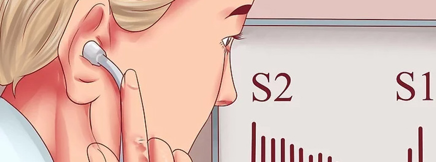
S1 and the 2nd heart sound (S2, a diastolic heart sound) are normal components of the cardiac cycle, the familiar “lub-dub” sounds.
S1 occurs just after the beginning of systole and is predominantly due to mitral closure but may also include tricuspid closure components. It is often split and has a high pitch. S1 is loud in mitral stenosis. It may be soft or absent in mitral regurgitation due to valve leaflet sclerosis and rigidity but is often distinctly heard in mitral regurgitation due to myxomatous degeneration of the mitral apparatus or due to ventricular myocardial abnormality (eg, papillary muscle dysfunction, ventricular dilation).
Clicks occur only during systole; they are distinguished from S1 and S2 by their higher pitch and briefer duration. Some clicks occur at different times during systole as hemodynamics change. Clicks may be single or multiple.
Clicks in congenital aortic or pulmonic stenosis are thought to result from abnormal ventricular wall tension. These clicks occur early in systole (very near S1) and are not affected by hemodynamic changes. Similar clicks occur in severe pulmonary hypertension. Clicks in mitral or tricuspid valve prolapse, typically occurring in mid to late systole, are thought to result from abnormal tension on redundant and elongated chordae tendineae or valve leaflets.
Clicks due to myxomatous degeneration of valves may occur any time during systole but move toward S1 during maneuvers that transiently decrease ventricular filling volume (eg, standing, Valsalva maneuver). If ventricular filling volume is increased (eg, by lying supine), clicks move toward S2, particularly in mitral valve prolapse. For unknown reasons, characteristics of the clicks may vary greatly between examinations, and clicks may come and go.
Diastolic heart sounds
Diastolic sounds include the following:
- 2nd, 3rd, and 4th heart sounds (S2, S3, and S4)
- Diastolic knocks
- Mitral valve sounds
Unlike systolic sounds, diastolic sounds are low-pitched; they are softer in intensity and longer in duration. Except for S2, these sounds are usually abnormal in adults, although an S3 may be physiologic up to age 40 and during pregnancy.
S2 occurs at the beginning of diastole, due to aortic and pulmonic valve closure. Aortic valve closure (A2) normally precedes pulmonic valve closure (P2) unless the former is late or the latter is early. Aortic valve closure is late in left bundle branch block or aortic stenosis; pulmonic valve closure is early in some forms of preexcitation phenomena. Delayed pulmonic valve closure may result from increased blood flow through the right ventricle (eg, in atrial septal defect of the common secundum variety) or complete right bundle branch block. Increased right ventricular flow in atrial septal defect also abolishes the normal respiratory variation in aortic and pulmonic valve closure, producing a fixed split S2. Left-to-right shunts with normal right ventricular volume flow (eg, in membranous ventricular septal defects) do not cause fixed splitting. A single S2 may occur when the aortic valve is regurgitant, severely stenotic, or atretic (in truncus arteriosus when there is a common valve).


plant of green extract