 Send a Whatsapp Message
 Free shipping on orders above £49 |
At a minimum, listen to the four basic auscultatory sites, first using the stethoscope’s
diaphragm and then the bell. Having a 3M® Littmann™ Stetoscope with tunable
technology allows you to hear different frequencies without repositioning the chestpiece.
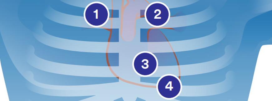
Base Right (Aortic area)
Base right (aortic) is the second intercostal space
to the right of the sternum. You can best hear
sounds from the aortic valve in this area.
Base Left (Pulmonic area)
Base left (pulmonic) is the second intercostal space
to the left of the sternum. You can best hear sounds
from the pulmonic valve in this area.
Lower Left Sternal Border
(LLSB) (Tricuspid area)
Left lower sternal border (tricuspid) is the fourth intercostal
space to the left of the sternum. You can best hear tricuspid
valve and right heart sounds in this area.
Apex (mitral) is the fifth intercostal space in the
midclavicular line. It’s easiest to hear mitral valve
and left heart sounds in this area.
Some of the best Cardiology Stethoscopes
-
 Littmann Stethoscope Master Cardiology Black / Brass FinishOriginal price was: £286.00.£259.00Current price is: £259.00.
 -
 Littmann Stethoscope Master Cardiology Black / Smoke FinishOriginal price was: £286.00.£259.00Current price is: £259.00.
 -
 Littmann Stethoscope Master Cardiology Black / Black EditionOriginal price was: £286.00.£259.00Current price is: £259.00.
The cardiac cycle consists of two periods: The first is a contraction (systole) and the second a relaxation (diastole). During systole, blood is ejected from the chambers of the heart and during diastole, the heart chambers fill with blood.
Ventricular systole causes closure of the mitral and tricuspid valves. Cardiac sounds are named according to the sequence of occurrence and are produced at specific points in the cardiac cycle.
The initial heart sound is called the first heart sound or S1. It occurs at the beginning of ventricular systole when the ventricular volume is maximal. The S1 corresponds to a point very early in the rise of the ventricular pressure curve where ventricular pressure becomes greater than atrial pressure and the mitral and tricuspid valves close.
This corresponds with the QRS complex on the ECG (electrocardiogram). On the graphic recording of heart sounds called a phonocardiogram, it is the first of the components recorded.
The second heart sound, or S2, occurs at the end of the ventricular systole, at the time of the diacrotic notch on the ventricular pressure curve. It is the second of the high frequency components recorded on a phonocardiogram.
The period between S1 and S2 represents ventricular systole.

