 Send a Whatsapp Message
 Free shipping on orders above £49
The most important aspect of lung auscultation is to be consistent in preparing and auscultating the
patient. This will allow for a complete assessment that is more accurate and increases the chances that
you will identify a subtle breath sound change. It is important to compare equivalent positions on each
side of the chest to identify asymmetry in the quality or loudness of the breath sounds.
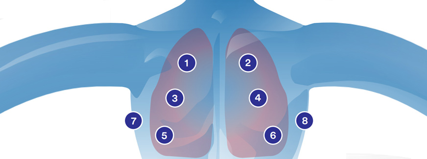
Knowing lung anatomy is important so that you can be sure that you auscultate at the correct positions.
The most common sites are shown below.
Optimal Environment
To optimise the effectiveness of auscultation the surroundings should be:
- Quiet – the ambient noise might interfere the heart and lung sounds.
- Warm -so that the patient feels comfortable while;the upper part of the body is being exposed. Also, it is to avoid shivering that may add the noise.
- Appropriate lighting – to allow good coordination between visual and auscultatory findings.
Respiratory Examination
Ideally, for Respiratory examination, the patient would be sitting.
Auscultate from side to side and top to bottom. Omit the areas covered by the scapulae.
- Usually the APEX of the lungs bilaterally (2cm superior to medial 1/3 of clavicle)
- Superior Lobes anterior (2nd intercostal space mid clavicular line) and posterior (Between C7 & T3)
- Inferior Lobes bilaterally anterior (6th intercostal space, mid-axillary line) and posteriorly (between T3 & T10)
- Middle lobe right anterior only (4th intercostal space mid-clavicular line)
Compare one side to the other looking for asymmetry and note the location and quality of the sounds you hear.
Technique
- Ask the patient to disrobe, as this will allow the stethoscope to be placed directly on the chest.
- Make sure the patient is sitting upright in a relaxed position, where this is possible.
- You should then instruct the patient to breathe a little deeper than normal through the mouth.
- The bell/diaphragm of the stethoscope is then placed against the chest wall.
- Auscultation of the lungs should be systematic, including all lobes of the anterior, lateral and posterior chest.
- The examiner should begin at the top, compare side with side and work towards the lung bases.
- The examiner should listen to at least one ventilatory cycle at each position of the chest wall.
- The examiner should identify four characteristics of breath sounds: pitch, amplitude, distinctive characteristics and duration of the inspiratory sound compared with the expiratory sound.


Its superb as your other posts : D, regards for posting.